|

|
In the area of thin-film nanomechanical analyses, Dr. Chang
investigated the mechanical properties, nanoscale responses and creep
behavior of metallic, dielectric, transparent conducting and protecting
films by using instrumented nanoindentations, as well as studied the
effects of residual stress and substrate on the mechanical tests of
thin films. Also, he tried to examine the stress-strain behavior,
early-stage dislocation activities and deformation mechanisms of thin
films by applying TEM observations and nanomechanics models. Through
the microscopic observations of different-grain-size Cu in the vicinity
of nanoindent marks, dislocation activities were clearly found in a
large-grain-size Cu, whereas grain boundary sliding and grain rotations
dominated the deformation of nanocrystalline Cu, suggesting the Inverse
Hall-Petch relation, which was published in Journal of Applied Physics
and evaluated as an outstanding study by the reviewer.
|
Left to right: deformation of different-grain-size Cu in the vicinity of nanoindent marks.
|
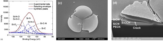
By using nanoindentations
and nanoscratch tests, Dr. Chang further studied the interfacial
delamination behavior and adhesion strengths of thin films, as well as
examined the effect of plasma treatments on interface chemistry
(bonding) and adhesion strengths. His findings contributed towards a
reliability enhancement of IC multilevel interconnects, and further
collaborations with the RD division at Taiwan Semiconductor
Manufacturing Company (TSMC) were accordingly conducted, yielding a
co-work patent of interfacial adhesion enhancement of thin dielectric
films. In addition, the nanomechanical properties and interface
adhesion of oxide, nitride, diamond-like carbon and quasicrystal films
were investigated under the collaboration projects funded by Industrial
Technology Research Institute (ITRI), Taiwan, as well as the
interfacial adhesion strengths of optoelectronic films measured under
the projects supported from semiconductor and optoelectronic industries
including ITRI, AUO Corp. and Rexchip Inc., etc.
|
Left to right: interfacial bonding configurations and interfacial delamination.
|
On the experience of
aforementioned thin-film and nanomechanical analyses, Dr. Chang began
the studies of the nanomechanical performance and deformation behavior
of biological tissues in recent years. Necessary analytical techniques
were built, and hard tissues (tooth and bone) were first investigated.
For the dental tissue, the mechanical properties of healthy teeth were
measured by nanoindentations, and the influences of bleaching agent,
soft drinks and Streptococcus Mutans on their microstructure and
mechanical properties were investigated. For the bone tissue, the
co-work (with the Department of Life Science, NCHU) on the effect of
osteoporosis on the microstructure, mechanical performance and
deformation/fracture behavior of bone was conducted, and the very
effective inhibition of osteoporosis by fermented milk was discovered.
The experimental results about the nanomechanical analyses of
biological tissues have been turned into several publications in
scientific journals including Journal of Materials Research, Journal of
The Mechanical Behavior of Biomedical Materials and Osteoporosis
International. Currently, Dr. Chang has begun attempts on the analyses
of the nanomechanical properties and deformation behavior of soft
tissues such as red blood cells and cytoskeleton.
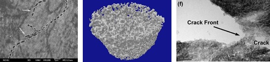 |
Left to right: dental enamel with S. Mutans, osteoporotic bone, and cracks in bone.
|
In recent two years, by the
funding supports from NSC, Taiwan, Dr. Chang further in-situ observed
the nanomechanical responses and deformation behavior of bone
nanostructure and nanoparticles/nanopillars under
nanoindentation/compression in a TEM. On the experience of
nanomechanical test, he has been capable of uniformly cutting
nanopillars (tip diameter < 70 nm), precisely manipulating a probe
in a TEM as well as in-situ observing the nanoscale deformation
behavior of nanomaterials under nanoindentation/compression in a TEM.
His recent studies revealed retarded crack propagations in a healthy
bone tissue by the bridging of collagen fibers and the distortions of
hydroxyapatite nanocrystals but a catastrophic fracture of osteoporotic
bone caused by rapid crack propagations and nanocrystal movements,
which was published in a top scientific journal Nano Letters. Moreover,
Dr. Chang’s recent findings in the in-situ deformation analysis of
single-crystalline Ag nanoparticles (size ~ 20 nm) in a TEM included
the ultrahigh strength of the nanoparticles and the remaining of
perfect lattice structure without dislocation activities. Furthermore,
the in-situ nanoscale deformation analyses of
single-crystalline/nanocrystalline/nanotwinned Cu,
metallic/ionic/covalent materials and unitary/multi-component materials
(high-entropy alloys) in a TEM have been carried out.
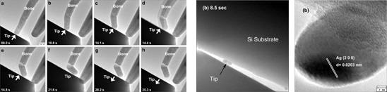 |
Left to right: in-situ nanoscale deformation of bone nanostructure and nanoparticle.
|
Dr. Chang further developed
an innovative route for spontaneously growing one-dimensional oxide
nanocrystals on oxide films in an ambient atmosphere, with the
application of mechanical stresses rather than the use of any
precursors or chemical solutions. Beyond conventional vaporous or
aqueous synthesis methods, a BHR (bond
breaking-hydrolysis-reconstruction) mechanism was proposed to elucidate
the stress-induced growth of oxide nanocrystals, and findings published
in Journal of Materials Chemistry. A mechanical way for stress-induced
graphitization of amorphous carbon was also developed, and the phase
transformation under nanocompression was examined in-situ in a TEM,
which lately yielded a publication in Carbon.
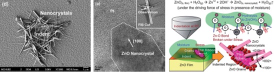 |
Left to right: stress-induced growth of ZnO nanocrystals and proposed BHR mechanism.
|
|
|
 |
|
|